

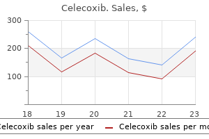
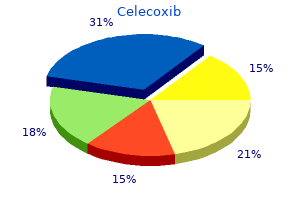
Celecoxib
F. Christopher Holsinger, MD, FACS
- Associate Professor, Department of Head and Neck Surgery
- Director, Program in Minimally Invasive and Endoscopic Head and Neck Surgery
- University of Texas MD Anderson Cancer Center
- Houston, Texas
The prevalence of iron deficiency is much higher in women than in men because of the toll of menstruation and pregnancy on the iron stores of women degenerative arthritis in my back buy 100 mg celecoxib with visa. The expansion of the blood pool that occurs during adolescence also leads to low iron stores that may be further depleted as a result of inadequate dietary intake arthritis x rays pictures celecoxib 200 mg free shipping. The latter factor contributes to the iron deficiency state in many women arthritis pain in back celecoxib 100 mg online, even in affluent societies arthritis psoriasis medication order 200 mg celecoxib with visa, as they embark on pregnancy. Mechanisms exist to ensure that the total-body iron content is maintained within a defined range. In specific contrast with other body constituents, control of iron content is imposed by limiting its entrance into the body rather than by increasing the excretion of any excess. The normal metabolism of iron is strictly weighted in favor of ensuring adequate iron reserves even at the cost of iron overload, which may result in hemochromatosis with organ damage created by the tissue accumulation of elemental iron. Storage pools of iron in the form of ferritin and hemosiderin are present within the liver, spleen, and bone marrow. The disparity in the size of these stores in men and women is attributable to the previously mentioned demands of menstruation and pregnancy in women. The tiniest compartment of iron within the body is transport iron (7 mg), in which iron travels while linked to the transport protein transferrin. Although transport iron is the smallest compartment, it is kinetically the most active and turns over several times a day as iron is transported to its various destinations within the body. Transferrin picks up iron from the gastrointestinal cells and delivers it primarily to cells engaging in hemoglobin synthesis. Transferrin also picks up iron from the storage depots in the daily recycling of iron stores. This system of conservation and recycling of iron serves to provide a constant supply of iron for the needs (30 to 35 mg) of daily hemoglobin synthesis. Only a tiny fraction of iron (1 mg) is lost each day via the pathway of sweating and epidermal shedding from the gut and urinary tract; this minuscule amount can easily be replaced from the food in a normal diet. The normal diet in the United States contains approximately 15 to 30 mg of iron each day, with every thousand calories in the diet containing about 6 mg of elemental iron. Iron is present in food as a portion of the heme ring in meats and in a less easily absorbable form as ferric hydroxide complexes in other foods. The acid environment of the stomach and its enzymatic secretions emulsify ingested food and liberate iron for absorption within the small intestine; pancreatic secretions counter this acidic pH and help control excessive absorption of iron. Ingestion of reducing substances such as ascorbate or succinate enhances iron absorption because of their effect on iron valence. Other substances such as phytates in cereals, tannates in tea, antacids, and certain antibiotics (tetracycline) may complex with iron and thereby hinder its absorption. Maximal absorption of iron occurs in the duodenum and upper portions of the jejunum. Malabsorptive states or bypass of these areas by gastrojejunostomy may contribute to iron deficiency. In normal health, the body must guard itself against iron overload by absorbing only one tenth of the iron available in the diet. The mechanism whereby such a limitation or "mucosal curtain" is imposed on iron absorption is still not defined; it appears to be regulated by some aspect of dynamic iron turnover 856 because hemolytic anemias, ineffective erythropoiesis, and hypoxemic states all have increased iron turnover and are all associated with increased iron absorption. The mucosal curtain is lowered by imposing a limit on the amount of iron that crosses the gastrointestinal membranes. Whatever iron is not needed by the body is diverted into storage molecules within gastrointestinal mucosal cells; this iron is lost from the body as these cells are exfoliated during the normal cycle of cell turnover. In the presence of iron deficiency, the body can increase its absorption efficiency at least five-fold to compensate easily and rapidly for any deficiency. In iron deficient states, in which iron needs are exaggerated, little iron is diverted to the storage form and the majority of the absorbed iron passes directly through the cells for plasma transport linked to transferrin. Failure to lower this mucosal curtain properly is thought to be the explanation for iron accumulation in primary hemochromatosis; such patients continue to absorb iron even in the face of total-body iron overload. Increased iron absorption also occurs with pancreatic insufficiency because of absence of the pH alteration contributed by normal pancreatic secretions; absence of this restraint on iron absorption explains in part the iron overload that often occurs in chronic alcoholic states.
Because the sensitivity of the genitalia to androgens decreases onward from early in fetal development pantrapezial arthritis definition discount celecoxib 200 mg without prescription, the extent of any virilization is important arthritis pain worse during period 100 mg celecoxib buy overnight delivery. Fusion of the labia and enlargement of the clitoris with or without formation of a penile urethra are observed in women exposed to androgens during the first 3 months of fetal development (see Chapter 246) arthritis in back symptoms buy discount celecoxib 200 mg online. Significant clitorimegaly in the absence of other signs of sexual ambiguity and in the presence of other signs of virilization requires marked androgenic stimulation and strongly implicates an androgen-secreting neoplasm in the absence of a history of ingestion of exogenous steroids arthritis feet physical therapy generic celecoxib 200 mg mastercard. The development of the labia minora in postpubertal women indicates the influence of estrogens. Overt anomalies of the distal genital tract and especially any evidence of obstruction to the escape of menstrual blood should be sought in the remainder of the pelvic examination. Under the influence of estrogen the vaginal mucosa changes during sexual maturation from a tissue with a shiny, bright red appearance with sparse, thin secretions to a dull, gray-pink rugated surface with copious, thick secretions. The history and physical examination quickly differentiate among several causes of amenorrhea, regardless of the age of the patient (Table 250-4). The various disorders of sexual differentiation and the other peripheral causes are often apparent on inspection. Distal genital tract obstruction should be identified at the time of pelvic examination even if the specific abnormality is not obvious. Any sexual ambiguity indicates the need for chromosomal analysis and the measurement of 17alpha-hydroxyprogesterone to rule out congenital adrenal hyperplasia. Tuberculous endometritis, especially in younger women, may also lead to this disorder. Without hormonal measurements it may be impossible to distinguish among individuals with chronic anovulation, in whom hypothalamic-pituitary-ovarian function is insufficiently coordinated to produce cyclic ovulation, and those with ovarian failure, in whom in most cases the ovaries are devoid of oocytes. Still, it is generally possible to form some strong clinical impressions about the cause of the amenorrhea. It can be noted if the patient has absence of, incomplete, or complete development of secondary sex characteristics. The presence of excess body hair or galactorrhea may provide clinical evidence of the pathogenesis of the amenorrhea. This schema must be considered as an adjunct to the clinical evaluation of the patient. This test is of limited value, however, because almost half the young women with premature ovarian failure experience withdrawal bleeding in response to progestin. To ascertain if the outflow tract is intact, an orally active estrogen, such as 2. Although hypothyroidism commonly results in anovulation, amenorrhea occurs in only some hypothyroid women. This is the case because prolactin levels are increased by nonspecific stressful stimuli, sleep, and food ingestion. Prolactin levels may be elevated in as many as one third of women with amenorrhea. Hyperandrogenic women need not be hirsute because some have relative insensitivity of the hair follicles to androgens. Consequently, some clinicians prefer to measure circulating free testosterone levels. Testosterone levels of >200 ng/dL should lead to investigation for an androgen-producing neoplasm, most likely of ovarian origin. Gonadal failure may begin at any time during embryonic or postnatal development and may result from many causes (Table 250-5). Normally the ovaries fail at menopause when virtually no functioning follicles remain. However premature loss of oocytes prior to age 40 years may occur and lead to premature ovarian failure, possibly from abnormalities in the recruitment and selection of oocytes. Circulating gonadotropin levels increase whenever ovarian failure occurs because of decreased negative estrogen feedback to the hypothalamic-pituitary unit. Several pathologic conditions with dysgenetic gonads have elevated gonadotropin levels and amenorrhea. The term gonadal dysgenesis refers to individuals with undifferentiated streak gonads without any association with either extragonadal stigmata or sex chromosomal aberrations. Because individuals with gonadal dysgenesis have the normal complement of oocytes at 20 weeks of fetal age but virtually none by birth, this disorder is a form of premature ovarian failure.

They could be due to mutations of yet unknown genes specifically involved in sex differentiation arthritis care back exercises 100 mg celecoxib purchase with mastercard. Even if such is not the case rheumatoid arthritis yellow eyes best celecoxib 200 mg, it is important to assign gender as early as possible arthritis in upper neck cheap celecoxib 200 mg visa. Gender identity is established very early in life arthritis health purchase celecoxib 200 mg visa, certainly by the time that speech is established. Gender confusion because of indeterminant or wrong assignment of gender may lead to severe emotional disorders later in life. Three diagnostic clues are helpful: gonadal location, presence of a uterus, and karyotype. If no mullerian derivatives are present, male pseudohermaphroditism caused by testosterone defects or malformations should be considered. It is prudent to wait a few days to assign the gender until common causes of sexual ambiguity are investigated and the various issues have been thoroughly discussed with the parents. However, once gender has been assigned, there should be no ambiguity in the sex of rearing to avoid confusion of gender. Proceedings of the Xth International Congress on Hormonal Steroids, Quebec City, Canada. In Thakker R (guest editor): Genetic and Molecular Biological Aspects of Endocrine Disease. An excellent review of the clinical and molecular aspects of androgen insensitivity. Swerdloff Christina Wang the testis is a bi-functional organ serving as the site of sex steroid. Thus, the testis controls both sexuality and the perpetuity of the species (fertility). In addition, androgens and their metabolites (including estrogens) serve essential metabolic roles and may be important inducers and effectors of brain function in men. The discussion in this chapter focuses on the issues of male reproductive physiology and its disorders: androgen deficiency, sexual dysfunction, infertility, and androgen excess states. The components of this system function in an integrative fashion to control the concentrations of circulating gonadal steroids required for normal male sexual development and function, for androgen- and estrogen-mediated metabolic effects on critical end organs such as brain, bone, muscle, liver, skin, bone marrow, and for immune systems. The reproductive axis is also responsible for normal germ cell maturation and sperm delivery necessary for male fertility. The heterodimer is required for biologic activity; the subunits can be detected in serum and may be increased in certain pathologic conditions. The seminiferous tubules contain Sertoli cells and germ cells at various phases of maturation. There is a diurnal rhythm of both gonadotropins (in young adult men) with higher circulating levels in the early morning and lowest levels in the evening. Puberty is heralded by night-time pulsatile serum patterns before obvious increases are noted in the daytime. Testis Function the testis is a complex organ consisting of (1) seminiferous tubules containing Sertoli cells and germ cells in various stages of maturation and (2) the interstitium where the steroid-secreting cells (Leydig), macrophages, and blood vessels reside. This process involves a steroid acute regulatory protein essential for steroidogenesis in the gonads and adrenal glands. Testosterone is the principal male hormone secreted by the testes, with about 7 mg produced per day. Testosterone synthesis occurs in the human testes through either the delta 4 or delta 5 pathways (see. Whereas both the delta 5 (left) and delta 4 (right) pathways exist, the delta 5 pathway predominates in the testis. It also can serve as a precursor for estradiol in some tissues where it binds the estrogen receptors (alpha and beta) to induce estrogenic effects. The spermatogenic compartment consists of the Sertoli and germ cells and is intimately interactive with the interstitial compartment. The Sertoli cells bridge the entire space between the basement membrane and the lumen of the tubules (see.

A number of magnification techniques can be used to improve visualization rheumatoid arthritis reddit 200 mg celecoxib buy with amex, and vasoconstrictive agents occasionally will be helpful to define normal vessels that constrict to epinephrine from tumors or inflammatory changes that are not as vigorously constricted by epinephrine arthritis diet wine purchase celecoxib 100 mg without prescription. Renal arteriography is especially useful for defining the extent and type of fibromuscular dysplasia and is diagnostically helpful in differentiating other stenotic lesions such as arteriosclerosis arthritis medication dogs 100 mg celecoxib purchase fast delivery, arteriodissections arthritis neck pain treatment buy celecoxib 100 mg fast delivery, emboli, thromboses, various types of vasculitides, and effects of trauma. There are no well-accepted criteria for choosing patients for renal arteriography, but the technique should be considered in those in whom therapeutically useful information is reasonably expected: patients with moderately severe hypertension, especially if they are young and have renal bruits on physical examination, and patients in whom renal arteriography can help define the nature of suspected tumors or help in the differential diagnosis of specific types of vasculitis. A modification of traditional renal arteriography is intravenous digital subtraction arteriography. In this technique, radiocontrast material is injected into either the inferior or superior vena cava, and digital subtraction imaging and filming are done later when the radiocontrast material circulates over the renal arteries. The technique has the advantage of using a lower contrast material dose, but the disadvantage is that it is mainly useful for diseases of the main renal artery and not its subsequent segments. The principle of this technique is that a computer constructs images mathematically from multiple x-ray absorption measurements from different projections of the body. To enhance the renal image, the studies are usually done with the aid of contrast material, except in circumstances in which the nature of renal calculi is being evaluated. These protons can absorb energy at only a very specific combination of local magnetic strength and the applied radiofrequency. Once the applied radiofrequency is stopped, the protons (small magnets) return to their previous lower-energy orientation in the magnetic field. The process of changing magnetic vector orientation (relaxation) creates a small electric current that is interpreted by the receiving coils. The signal is dependent on unique magnetic surroundings of protons in tissue, and the ultimate image is constructed by complicated computer technology from evaluating the proton signals in three separate magnetic field axes. The resulting images from tissues of given characteristics form images of differing brightness. The organs are well outlined by differences in the magnetic environment of capsules and surrounding tissues. Nevertheless, it cannot differentiate the nature of renal masses such as abscess versus carcinoma. Radionuclide activity can be measured as counts per volume of fluid by appropriate counters or by number of energy emissions as detected by a gamma camera (alpha and beta emittors do not have long enough penetration to be of use clinically). In each case, an ideal tracer is one that is only cleared from the body by glomerular filtration. In this technique, stable blood concentrations of radionuclide must be achieved by either continuous infusion techniques or by single subcutaneous injections with suitable equilibration periods (usually 40 to 60 minutes). Although the technique is not easy, it is useful in situations in which collecting urine is difficult, such as in pediatric populations or patients with urinary diversions. Clinically, the most effective compound that is available is radiolabeled p-aminohippurate, which has an extraction efficiency of 87%. Other compounds have been developed with both lower and higher extraction efficiencies. The renal blood flow can be calculated from the clearance of radioactive nuclide from blood at short intervals up to 2 hours. The renal blood flow can be calculated from the rate of decrease of the compound from blood and corrected for the extraction efficiency. These are only rough estimates of renal anatomy but have some advantage in that allergic reactions or induced nephropathy is essentially non-existent to radioactive compounds as compared with radiocontrast materials. In this technique, radiopharmaceuticals that are taken up by the kidney are injected. Images are then collected at 30-second intervals for 2 minutes and then at 5-minute intervals. The technique is useful to compare the function of one kidney versus the other and gives a general outline of renal size and shape. One of the most important uses of radionuclides in nephrology is to measure the function of one kidney relative to that of the other when nephrectomy is considered. The technique is similar to imaging, but in this circumstance the uptake of radiopharmaceuticals by one kidney is compared with that of the other. It is important to start images only after adequate mixing of radionuclide has occurred in circulation-usually within 2Ѕ to 3 minutes.
A careful history arthritis in knee after meniscus removal celecoxib 200 mg cheap, physical examination medicine used for arthritis in dogs discount celecoxib 200 mg on line, and directed imaging and laboratory studies will reveal the probable infectious focus in most patients rheumatoid arthritis zero positive celecoxib 200 mg purchase without a prescription. However arthritis pain during period buy 200 mg celecoxib with mastercard, elderly, debilitated, and immunosuppressed patients may not exhibit the usual localizing clinical signs. In some patients, especially those with severe neutropenia, no site is identified. Second, patients usually manifest one or more signs of the systemic inflammatory response. Elderly patients may present with tachypnea-induced respiratory alkalosis and mental status changes as the only signs of sepsis. Third, septic patients may develop evidence of shock, such as hypotension, lactic acidemia, and progressive organ system dysfunction. The diagnosis of sepsis is confirmed by culturing pathogenic organisms from blood or from the likely site of infection. Blood cultures are positive in only 40 to 60% of patients with clinical manifestations of septic shock, probably owing to the intermittent nature of the bacteremia and the high incidence of prior antibiotic administration. A Gram stain from an abscess, empyema, or other usually sterile site can provide invaluable early diagnostic information. First, the infection site can be eradicatedwith antimicrobials, surgical drainage, or both. Second, the serious disturbances in cardiovascular, respiratory, and other organ system 511 Figure 96-3 Algorithm for diagnostic evaluation and management of sepsis and septic shock. Shock secondary to sepsis is a very major disease that should be treated aggressively. When the diagnosis is seriously entertained, blood cultures (usually three) and cultures of relevant body fluids and exudates should be obtained rapidly. Several large retrospective trials have provided convincing evidence that early appropriate antimicrobial therapy. A broad-spectrum regimen with activity against gram-positive and gram-negative organisms should be chosen. Generally, drugs should be administered intravenously at maximum recommended dosages, and bactericidal agents are preferred over bacteriostatic agents. Many physicians favor using at least two effective antimicrobial agents in neutropenic patients with gram-negative pneumonia and a two-drug synergistic combination when treating serious enterococcal infection (see Chapter 314). Anaerobes are likely pathogens in intra-abdominal infections, aspiration pneumonia, and abscesses. Intravascular catheter infection should raise the possibility of methicillin-resistant staphylococcal infection and the need for vancomycin therapy. In up to one third of patients, especially those who are neutropenic, no organism or source will be identified. Such patients require a broad-spectrum regimen effective against gram-positive, gram-negative, and anaerobic organisms such as (1) vancomycin, gentamicin, and metronidazole or (2) ceftazidime and gentamicin. The need for early antifungal therapy with amphotericin B should be considered in neutropenic, immunosuppressed patients and in those unresponsive to antibacterial regimens. Before the general availability of intensive care units, gram-negative bacteremic shock had a higher than 90% mortality. Now, about 50% of such patients survive, largely because of treatment in intensive care units, in which cardiac rhythm, blood pressure, cardiac performance, oxygen delivery, and metabolic derangements can be monitored and abnormalities can be corrected. Although no prospective trial has evaluated outcomes with and without intensive care unit support, two retrospective studies have reported a significantly reduced mortality in septic shock when patients were managed with aggressive hemodynamic support by critical care personnel. A controlled, prospective trial of intensive care unit support has been conducted in dogs with gram-negative sepsis; survival was increased only in the animals that received both antibiotic therapy and cardiovascular support. Patients with septic shock who remain hypotensive after a 1- or 2-L volume resuscitation should have arterial and pulmonary artery catheters placed to allow serial evaluations of blood pressure, ventricular filling pressures, cardiac output, and oxygen delivery. Initial emphasis should be placed on restoring mean blood pressure to greater than 65 mm Hg.
Purchase celecoxib 100 mg otc. Pathogenesis of rheumatoid arthritis.
References
- Branca P, McGaw P, Light R: Factors associated with prolonged mechanical ventilation following coronary artery bypass surgery, Chest 119:537-546, 2001.
- Akhter M, Lajos TZ: Pitfalls of undetected patent foramen ovale in off-pump cases, Ann Thorac Surg 67:546, 1999.
- Ghatak NR, McWhorter JM. Ultrastructural evidence for CSF production by a choroid plexus papilloma. J Neurosurg 1976; 45:409-415.
- Steed DL, Brown B, Reilly JJ, et al: General surgical complications in heart and heart-lung transplantation, Surgery 98:739, 1985.
- Kelly WD, Merkel FK, Markland C: Ileal urinary diversion in conjunction with renal homotransplantation, Lancet 1(7431):222n226, 1966.
- Oren I. Breakthrough zygomycosis during empirical voriconazole therapy in febrile patients with neutropenia. Clin Infect Dis. 2005;40(5):770-771.