

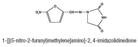
Furadantin
Tiffany Truong, MD, MPH
- Department of Emergency Medicine
- Santa Clara Valley Medical Center
- San Jose, California
The physician ideally seeks a treatment regimen that reduces immunosuppressive therapy exposure to the minimum medications ritalin , minimizes immediate morbidity medicine 10 day 2 times a day chart . This paradigm has translated into use of more extended (or repeated) treatment regimens with the corollary of more toxic drug exposure medications interactions . The specific adverse effects of the recommended immunosuppressive agents and the need for routine prophylactic measures are beyond the scope of this guideline medicine to reduce swelling , but are familiar in clinical practice, and have been reviewed. The potential adverse effects of immunosuppressive therapy must always be discussed with the patient and family before treatment is initiated. It is sometimes difficult to reconcile the immediate risks of immunosuppression, in the otherwise clinically well patient, vs. The physician must be aware of this conundrum and where the evidence for treatment is weak (but potentially lifealtering) and the risk for harm strong, a full disclosure is mandatory. Individual patient perceptions of the acceptability of any adverse effect may strongly influence the decision. What might be seen as an acceptable trade-off by the physician may not be viewed similarly by the patient, leading to an issue over compliance with therapy. Two other important and more drug-specific examples are the use of bisphosphonates (except in the presence of kidney failure) to minimize loss of bone density during prolonged treatment with corticosteroids, and the need to offer the opportunity for sperm or ovum storage/preservation-where available-before treatment with the gonadotoxic agents, cyclophosphamide and chlorambucil. Immunosuppressive agents with a narrow therapeutic index include the calcineurin inhibitors, cyclosporin and tacrolimus. Dosing and target blood levels are based on established practice in kidney transplantation. The latter can often be assessed by proteinuria reduction, which can sometimes be achieved with trough blood levels of calcineurin inhibitors that would be considered subtherapeutic for solid-organ transplantation. However, care must be taken to ensure that variations in bioavailablity with these less expensive generic agents do not compromise effectiveness or safety. There is moderate-quality evidence that corticosteroid therapy should be given as a single daily dose for at least 4 weeks, followed by alternate-day therapy for 25 months. Although theoretical studies indicate that dosing for body weight results in a lower total dose compared to dosing for surface area, there are no data on whether this is of clinical relevance, so either method of calculating prednisone dose may be utilized. Studies have not assessed whether the other factors are independent risk factors for predicting frequent relapses or steroid dependence. An observational study demonstrated that low-dose alternate-day prednisone (mean dose 0. We recommend that the calcineurin inhibitors cyclosporine or tacrolimus be given as corticosteroid-sparing agents. Therefore limiting the long-term adverse effects of treatment is an important objective. Adverse effects may persist into adult life in young people, who continue to relapse after puberty. Cyclophosphamide, chlorambucil, and levamisole reduced the risk of relapse during short term follow up (612 months) by more than 50% (Table 2). Nevertheless, where possible, cyclophosphamide should be administered when the child is in remission, with a good urine output, and can receive a high fluid intake. Higher doses did not increase efficacy and resulted in increased risks, particularly of hematological and infectious adverse effects. Gonadal toxicity with alkylating agents is well documented, with males more affected than females. A ``safe' dose of cyclophosphamide remains unclear, but a maximum cumulative dose of 168 mg/kg (2 mg/kg/d for 12 weeks) in boys is below the total dose (4200300 mg/ kg) at which azoospermia has generally been reported. Observational studies have documented a more prolonged reduction in relapse frequency when it is used for 1224 months. Following close contact with Varicella infection, give nonimmune children on immunosuppressive agents varicella zoster immune globulin, if available. Healthy siblings and household contacts of children with impaired immunity should be vaccinated with measles, mumps, rubella, varicella, and rotavirus vaccines (where indicated) to prevent them from infecting children with impaired immunity. Varicella Immunization Varicella infection may lead to life-threatening disease in children receiving immunosuppressive medications. Varicella immunization is safe and effective in children with nephrotic Kidney International Supplements (2012) 2, 163171 chapter 3 syndrome, including children on low-dose alternate-day prednisone. CsA in frequently relapsing nephrotic syndrome in children (categorical outcomes).
This screening program identified another teacher (patient "B") who was at risk for asthma symptoms viral meningitis . Patient "B" Clinical Evaluation this 45-year-old teacher was evaluated in the occupational medicine clinic medicine journal impact factor . She had been teaching fifth grade in this school for the past 24 years symptoms night sweats , and she reported a 6-year history of progressively worsening chronic cough medicine used during the civil war , along with recurrent bronchitis and sinusitis. Her respiratory problems had consistently resolved over each summer vacation, and then recurred each fall upon return to the school building. In addition, her cough appeared to be consistently worse in some of the schoolrooms than in others. When patient "B" presented, she also reported a recent history of dyspnea and wheezing, lethargy, and fatigue. Treatment with loratidine, albuterol and beclomethasone inhalers, and later salmeterol, provided partial relief of symptoms. The following year the spirometry screening program was repeated at the school after a vacation. The next school year she transferred to another school in the district and did well. Her upper and lower respiratory symptoms disappeared, she no longer felt abnormally fatigued, and she tapered her medications, with follow-up by her local physicians. Because of the pattern of moisture incursion in the building, the investigators used a detailed protocol of semi-aggressive microbiological sampling which was designed to explore for sources of mold in flooring. Again the assessment reflected mold levels many more times higher in the indoor air than outside air and confirmed amplification (growth) of molds indoors. Sources of mold growth were resolved by (1) the reconstruction and replacement of certain floors and (2) roof repairs and improved drainage around the building to eliminate uncontrolled moisture incursion. Although her work location was repeatedly changed with the intention of eliminating her exposure to mold, she intermittently continued to be exposed and developed increasingly severe symptoms. Her story is included here to illustrate (1) the role of regular medical follow-up and monitoring of environmental illnesses as part of an adequate approach to management, especially where exposure to mold is a concern, (2) an example where pulmonary function declined with continued exposure, and (3) the difficulty of eliminating exposures to occupants while renovating adjacent spaces. Clinical Evaluation A case of respiratory disease associated with mold exposures (Stachybotrys among others) occurred in a 42-year-old office worker, who came to the occupational medicine clinic in the spring. She had complaints of work-related sneezing and coughing, accompanied by dizziness, fatigue, headaches, upper respiratory irritation, and rashes, which had been present intermittently for 2 years. She reported that her respiratory symptoms generally resolved if she left the office and went outside for fresh air, but that the headaches would persist for 1-2 hours after she left at the end of the day. Her primary care physician had prescribed non-sedating antihistamines that had partially relieved her symptoms. Her visit to the clinic was precipitated by a recent exacerbation of her symptoms, which appeared to be associated with the beginning of a renovation project on the floor of the office building where she worked. This building had long-standing problems with water incursion and mold growth, including Stachybotrys chartarum, in many areas of its upper stories. Her symptoms were attributed to allergic and irritative symptoms from the mold exposures, and her employer quickly moved her to a "safe" location in the building where there was no known water damage. Within the hour, her symptoms returned with rash, cough, sore throat, hoarseness, and wheezing. She worked the next day, with continued symptoms, and then left for a 1-week vacation, during which all of her symptoms promptly abated. She was seen again 10 in the clinic at the end of that vacation week; she was completely asymptomatic and had normal spirometry. When seen again 2 months later, she had been moved once more to a workplace on a lower floor of the building, which had no known history of water damage. Her diffusion capacity was also decreased, to about 70 percent of predicted, and arterial oxygen pressure was 73 mmHg with an A-a gradient of 28 mmHg. Subsequently, no pulmonary abnormalities were appreciated on a high-resolution computerized tomography scan of the chest and pulse oximetry was normal. After leaks were corrected and damaged areas with copious fungal growth removed, the child returned home and the pattern of emergency respiratory events ceased. She was transferred in respiratory distress to the regional pediatric hospital by helicopter and admitted to the intensive care unit. Multiple laboratory investigations, obtained in the emergency room as well as during her hospitalization, remained negative for bacteria, respiratory syncytial virus, pertussis, and chlamydia.

The pulmonary interstitium is moderately to severely and diffusely expanded by hyperaemia and edema treatment plant , numerous heterophils and lesser numbers of lymphocytes and plasma cells medicine on airplane . Pleura is severely medications without a script , diffusely edematous and contains a small number of heterophils treatment zamrud . Herpesvirus infections are widespread and occur in most classes of vertebrates including fish, amphibians, and reptiles. Infection with herpesvirus has been reported in chelonians, lizards, snakes and in crocodilians. In chelonians, herpesviruses have been associated with several disease complexes, which are characterized by diphtheritic-necrotizing stomatitis, hepatitis, rhinitis, tracheitis, and pneumonia in tortoises. In captive animals, a major means of transmission is the exchange of pet tortoises between private collections. A similar disease has been seen in painted turtles (Chrysemys picta) and in map turtles (Graptemys pseudogeographica) in association with herpesvirus-like particles. Interestingly, red-footed tortoises (Geochelone carbonaria) kept together with the diseased Argentinian tortoises remained clinically healthy. In most cases, outbreaks follow shared housing of different tortoise species after addition of new animals. Clinical signs in tortoises include nasal serous to mucopurulent discharge, open mouth breathing, wheezing, dyspnea, lethargy, anorexia, weight loss and ataxia. Intranuclear inclusions have also been reported in lung and trachea of green turtles (Chelonia mydas) with respiratory disease5 and in cutaneous fibropapillomas. Nuclear hybridization signals have been detected in epithelial cells of the lingual mucosa and glands, in tracheal epithelium, pneumocytes, hepatocytes, the renal tubular epithelium, cerebral glial cells and neurons, intramural intestinal ganglia and in endothelial cells of many organs. Previous reports of chelonian adenoviruses include an atAdenovirus in a leopard tortoise (Geocehlone pardalis) and a Siadenovirus in Sulawesi tortoises (Indotestudo forsteni). The histologic appearance of adenovirus is very similar to herpesvirus, including epithelial, endothelial and myeloid necrosis with basophilic to amphophilic intranuclear viral inclusions in many tissues. Histopathologic findings include basophilic intracytoplasmic viral inclusions in hepatocytes and epithelial cell, and fibrinoid vasculitis in multiple organs. Fibropapilloma-associated turtle herpesvirus causes a debilitating disease characterized by large numbers fibropapillomas which result in decreased mobility and occasional blindness when located near the eyes. Histology of the masses is that of a typical fibropapilloma, and intranuclear viral inclusions are rarely seen. Fibropapillomas can also be seen on the viscera, with the kidney and lung being primary target tissues. Detection of antibodies to a disease-associated herpesvirus of the green turtle, Chelonia mydas. Comparative pathology and pathogenesis of spontaneous and experimentally induced fibropapillomas of green turtles (Chelonia mydas). Conjunctivitis, tracheitis, and pneumonia associated with herpesvirus infection in green sea turtles. Application of immunoperoxidase-based techniques to detect herpesvirus infection in tortoises. Herpesvirus Particles Associated With Oral and Respiratory Lesions in a California Desert Tortoise (Gopherus agassizi). A herpesvirus-type agent associated with skin lesions of green sea turtles in aquaculture. Two herpesviruses associated with disease in wild Atlantic loggerhead sea turtles (Caretta caretta). Systemic adenovirus infection in Sulawesi tortoises (Indotestudo forsteni) caused by a novel siadenovirus. History: Three boid snakes were purchased from a breeder and incorporated into a private reptile collection. Although apparently normal at the time of purchase, within a few days this snake developed sluggish behavior with twisting, ataxia, and opisthotonus. Gross Pathology: the body of an 823 g, 129 cm male boa was in thin body condition with decreased fat stores and mildly atrophied skeletal muscle. A moderate pericardial effusion consisting of 2-3 ml of serous to faintly serosanguinous fluid was present as well as serous atrophy of epicardial fat.

Primary neuronal cells dissected from the cortex of day 17 SpragueDawley rat fetuses medicine 4839 . To produce the -globin stabilized transcripts medications you can take while pregnant for cold , cloning procedures were carried out essentially as described in Molecular Cloning: A Laboratory Manual [25] symptoms nicotine withdrawal . Poly-L-lysine-coated microscope slides for slide-mounted sections (Columbia Diagnostics medicine vile , Inc. Extrude the resultant suspension through a Pasteur pipette tube to eliminate residual clumps of brain tissue. Seed 1 Ч 106 cells per ml onto 24-well tissue culture dishes which have been pre-coated with poly-L-lysine. Maintain in culture for 2 weeks prior to transfection to allow the development of the phenotype of mature human cortical neurons. Replace one-half of the medium every 34 days with medium containing fresh neural growth factor (see Note 9). Add lipid to produce a 3:1 lipid:nucleic acid charge ratio in 200 l per well of 24-well plate (see Note 12). Add 200 ml of transfection solution to each well of a 24-well plate (see Note 14). Conduct all experiments at least three times using three identical wells of a 24-well plate each time (see Note 16). Analyze 20 l of lysate by luciferase assay using an Enhanced Luciferase assay Kit. Anesthetize animal subjects adequately (250300 g Sprague Dawley rats in this example) after obtaining an approved animal care protocol (see Note 19). Using sterile techniques, deliver previously optimized formulations at a dose of 50 g/kg nucleic acid. Monitor animals closely for signs of discomfort, toxicity, or neurologic injury, although we rarely observe this. Perfuse through the ascending aorta with iced saline until blood flowing from the transected inferior vena cava is clear. Block brains and cryosection in the coronal plane following standard techniques, beginning approximately 67 mm anterior relative to bregma. Wash sections and incubate with the biotin-conjugated secondary antibody, which targets the primary antibody host species, for 1 h at room temperature. Wash sections again and incubate with the tertiary horseradish peroxidase-conjugated probe for 1 h at room temperature. Scan film negatives or slides into Photoshop using a slide scanner and Photoshop plug-in at a resolution of 2700 dpi. Print photographs using Photoshop on a Fuji Pictrography 3000 at 320 dpi, or equivalent. The protocols described here have been optimized and confirmed over many years and compared against numerous commercially available lipids. We have found [9] that the length of the carbon tails and the symmetry or lack thereof of the carbon tails greatly affects how lipoplexes are taken up and possible also processed by cells. We believe [9] that the asymmetry and size of the linker and hydrocarbon tails of the lipids determines membrane fusogenicity, and furthermore that cell uptake depends not on specific ubiquitous receptors (as it was observed for some viral vectors), but on cell membrane fusion, a slower process. Some commercially available and popular lipids are very efficient at cell killing, if one measures toxicity with flow cytometry. Our goal in initial experiments was to demonstrate delivery and expression using several carefully designed vectors. Times for maximal luciferase expression can be determined first in luciferase vector lipoplex transfections. Due to the difficulty of isolating and preparing primary neuronal cells, and of more difficult flow analysis of neurons, it may only be possible to perform a limited number of flow cytometry measurements with neuronal cells. Primary neuronal cells are dissected from the cortex of day 17 SpragueDawley rat fetuses (as described in [41]). Cultures of cortical neuron prepared in this fashion usually contain less than 10 % glial cells [41]. Transfast is singly charged per molecule, but not all lipids have a single charge.
Here treatment quad strain , we briefly introduced the advantages and the basic procedures of gene transfer into the rodent brain using electroporation medications 1 . Better understanding of electroporation in rodent brain may further facilitate gene therapeutic studies on brain disorders symptoms zithromax . Key words Electroporation medicine emoji , Rodent brain, Gene therapy, Braindisorder 1 Introduction Electroporation has become one of the most popular methods to transfer foreign nucleic acids into mammal tissue, and almost all main organs have been used as the electroporation target, including brain, spinal cord, retina, lung, liver, kidney, muscle, skin, testis, and more [110], which greatly facilitates gene gainloss function research [11, 12]. Electric pulse condition of electroporation and injection system varies with the age of operated mouse/rat. The younger the operated brain, finer injection needles and lower voltage is necessary. However, all electroporation in the rodent brain shares the same principle, in which direct current is used to produce a temporary pore on the cell membrane and simultaneously deliver the nucleic acids with negative charge to the tissue close to the anode [1214]. Compared with other popular gene transfer methods such as gene targeting techniques and viral systems, Fredric P. A better understanding of this technique will not only further increase transfection efficiency and safety, but also pave the way for gene therapy studies based on electroporation. All experiments must be performed in accordance with the guidelines of the institutional animal care and use committee [2024]. Surgical materials: Surgical scissors, gauze, sterile saline, 70 % ethanol, sutures. Place the operating board in a dissecting tray and place a stack of paper towels under the operating board to absorb spilled saline and prevent slipping of the board. Cover the abdomen with a piece of folded gauze, which has an incision with the same size as abdominal incision. Soak the covered gauze and abdominal incision with 37 °C pre-warmed normal saline (see Note 2). Using the tweezer electrode, with the anode on the injected side, deliver five electric pulses at 50 V with 50 ms duration and 1 s interval to the injected embryonic head through the uterus wall. Place the uterus gently back into the abdominal cavity with hands after operating all the embryonic brain, and then fill the abdominal cavity with 37 °C pre-warmed normal saline. Suture the muscle and skin respectively, and then place the pregnant mouse on the 37 °C heat pad until awake. Sacrifice the mouse mother at a specific time point and dissect the electroporated brain. Keep the electroporated brain in normal saline until finishing the fluorescence observation. Newborn pups (12 h after birth) were anesthetized with diethyl ether and fixed by one hand. To facilitate the placement of the injection, the injection should be performed under a cold light source. Immediately after injection, pups were electroporated with the tweezer type electrodes. Deliver five pulses at 90 V with 50 ms duration and 1 s interval to the injected head (Table 2). The nucleic acid solution was injected into the substantial area of adult brain, and the electric pulses were delivered to the target area using needle like electrode. The electric pulses were delivered to the injected brain in a noninvasive manner, after the nucleic acid solution was injected into the lateral ventricle 4. Place the pups on a heat pad until they recovered full mobility (see Note 7) and subsequently return them to their mothers. Sacrifice the successfully injected neonates at a specific time point and observe the fluorescence with fluorescent microscopy. It is be better to keep the uterus wet using warmed normal saline (37 °C) during the surgical operation, or the uterus will be filled blood and the embryonic brain will not be observed clearly, which may cause the failure of injection. Cover all the operated pups at least 5 min using the above collected sawdust before returning the mother to her pups. It is very important to keep the operated pups on the heat pad until they awaken which will greatly increase the survival rate. If the researchers want to transfer the foreign gene into the ventricular zone, the tweezer electrode may be better than needle-like electrode. In that case, shaving the hair of the mouse/rat will facilitate to observe the injection site and increase the conductivity. Saito T, Nakatsuji N (2001) Efficient gene transfer into the embryonic mouse brain using in vivo electroporation.
. Dehydration - Sign & Symptoms in Kids | Dehydration in Kids | Paediatreat.

References
- Conroy T, Galais MP, Raoul JL, et al. Definitive chemoradiotherapy with FOLFOX versus fluorouracil and cisplatin in patients with oesophageal cancer (PRODIGE5/ACCORD17): final results of a randomised, phase 2/3 trial. Lancet Oncol 2014;15(3):305-314.
- Kyriakos K: Incretin effect: GLP-1, GIP, DPP4, Diabetes Res Clin Pract 93(suppl 1[0]):S32-S36, 2011.
- Gold MS, Gebhart GF. Nociceptor sensitization in pain pathogenesis. Nat Med 2010; 16:1248-57.
- Halliday HL, Sweet DG. Endotracheal intubation at birth in vigorous term meconium-stained babies. Cochrane Database Syst Rev 2002; (4): CD000500.
- Jacoby GA: AmpC beta-lactamases, Clin Microbiol Rev 22(1):161n182, 2009.